

Use of a Sling in Non-ambulatory Long-legged Birds
- Sep 10, 2021
- 6 min read
The management of avian patients who are non-ambulatory is difficult at the best of times, but the management of long-legged species - cranes, storks, stilts, herons etc. - is further complicated by their particular anatomy. These patients can develop severe irreversible myopathy if left to be recumbent for even relatively short periods of time, including during sedation or anaesthetic procedures of only 1-2 hours. In addition, a bird of this type that finds itself unable to move around away from threats can further injure itself by thrashing around in an attempt to stand. Given the delicate nature of their long leg bones, fractures are not uncommon in the hospitalised patient.
The use of a sling to support bodyweight and restrict movement is a well-documented therapy in the avian veterinary field in a wide range of species.


Below is a summary of two cases of long-legged avian patients seen at the NZCCM where a sling was used as part of the therapeutic plan and inpatient management.
Case 1 “Liyongo”, a 5-month-old Greater Flamingo Phoenicopterus roseus, presented to the veterinary team for assessment of sudden onset of lameness of the right leg. After 24 hours

of rest, pain relief and stabilisation, he was anaesthetised for a thorough examination, blood tests and X-rays. X-rays showed a very small fracture on one side of the growth plate at the top of the right tarsometatarsus bone. The growth plate is the area at the end of each bone in which bone is formed and grows in a young animal. Growth plates fuse as the animal reaches adulthood when the bones are at full length. Fractures of growth plates can be corrected surgically, but surgical complications may lead to growth deformities in this bone. Splinting is also tricky for the same reasons – any restriction of the joint for too long can prevent the limb from growing in a natural shape.
Figure 1:Liyongo on Day 1 of presentation
In a domestic animal, any treatment plan for a growth plate fracture would include strict restriction of movement, such as keeping the animal in a small crate or pen and ideally preventing any bodyweight from being supported by that leg.
Whilst we were able to begin all other aspects of the treatment plan, such as anti-inflammatories, profound pain relief, fluids and tube feeding, the patient would become stressed when handled and kick out, risking further injury and potentially worsening the growth plate fracture.

The correct way to restrain a long-legged bird is to hold the tibiometatarsus (“hock”) joint in a 3-fingered hold in one hand whilst the rest of the bird’s body is tucked under the handler’s other arm, facing backwards. Doing this gives control of the legs, preventing them from crossing and tangling or kicking out and connecting with a solid object which could cause injury to the leg. As the “hock” was the fracture site, it was not suitable to restrain the patient in this way. In addition, he was now spending a lot of time sitting down on his hocks and was finding it more and more difficult to stand due to the periods spent sitting.
Figure 2: Correct restraint of flamingo for veterinary examination
Liyongo was placed in a sling to restrict the bird’s movement and take the weight off the leg.

Figure 3: First version of the flamingo sling, incorporating wing restraint. Note mirror in the enclosure to provide "company"
Initially, this was a simple towel and strap arrangement hanging from an overhead beam. Particular care was taken to cut holes in the towel to allow the legs to pass through and hold the body comfortably. The leg holes needed to be large enough so that they were not tight around the tops of his legs, and the angle of the sling needed to be such that the body was supported in a natural position with the front of the body slightly higher than the rear. Care had to be taken to ensure that no pressure was placed on the crop area. The sling was placed at a height that allowed the bird to just place his feet flat on the floor and allow a slight flex of his hocks. If the sling were too high, preventing contact with the ground, he would have panicked and kicked out. If it were too low, it would not have been supporting his body weight.
The first version of the sling met all the requirements needed; however, the flamingo could get his neck or wings caught around the strapping, and there was a risk that he might be able to manoeuvre himself partway out of the sling and then fall and become entangled. A range of different options was debated, and our works department manufactured sling 2:0.

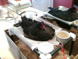
The sling comprised of a square wooden frame, large enough that no part of the flamingo could reach the frame when settled in the sling. A soft fleece blanket was stretched across the frame and fixed to each side, and leg holes were cut as previously in the blanket. The blanket had enough stretch to sag gently when the bird’s body was placed in it and
was adjusted as previously so it would be at the correct height. Care was taken to ensure a small folded towel was placed under the cloaca to be replaced when soiled, and this was checked frequently to ensure it was not bunching and causing discomfort.
Figure 4: Sling 2.0 demonstrating support of full bodyweight when required

Although the new sling served the intended purpose well, the flamingo remained agitated and initially would try to jump out over the frame. Sheets were hung to create a fabric “box”, enclosing the sling so that he could not see out.
Eventually, a constant low-dose infusion of IV sedative was needed to keep the bird calm, alongside the usual considerations
such as dimming the lights, keeping noise to a minimum and minimising the number of times the room was entered. Tube feeding was achieved by simple restraint of the head and restricting wings, and we felt the prognosis was hopeful.
Figure 5: Feet can be placed flat on the floor with slightly flexed hocks
Sadly despite our best efforts, the demeanour of the patient deteriorated over the course of the fortnight, and eventually, the decision was made to euthanase on welfare grounds. Flamingos are a strongly social flock species. Although we tried to mitigate his solitary hospitalisation with mirrors and life-size flamingo posters placed around the walls, this was a 5-month-old chick who had never been separated from his flock was not comfortable in the presence of humans. “Liyongo” was an interesting case that gave us a lot of learning opportunities. We have reviewed this and plan for different strategies that could be attempted in future cases.

Figure 6: A difficult case to manage
Case 2
“Mrs Brolga”, a 27-year-old female Brolga crane Grus rubicunda, was presented to the

Veterinary team with a history of being subdued and not eating for 24-48 hours. On initial approach, this bird was found sitting down without attempting to stand, which is unusual for this species. A differential diagnosis of myopathy secondary to trauma or over-exertion was presumed and confirmed on blood test results. By the following morning, despite medications, fluid therapy, and supportive care, the bird could not raise off her hocks unaided. A sling was fashioned to support her weight and keep her legs in a natural (slightly flexed) position rather than folded under her. She was placed in the sling for periods of about one hour up to three times daily and otherwise had a deep straw bed to lie down on.
Figure 7: Mrs Brolga in a sling
During her time in the sling, her legs were gently manipulated in range-of-motion exercises. If she became agitated by our presence, a hood was placed over her head to keep her calm.

She was force-fed with pieces of her usual diet and given liquids and liquid food by stomach tube. By the end of day three of treatment, she could walk and remain standing after a session of being in the sling. She steadily improved over the next few days, walking independently, starting to eat by herself, and resist capture. She remained on treatment for a few more days but eventually made a full recovery
Figure 8: Mrs Brolga having physiotherapy while in the sling. Note the use of a hood to keep the patient calm
Author’s notes
Both of these cases demonstrate the dynamic requirements when treating wild birds. The initial treatment plan for any animal must be constantly re-evaluated and altered for the patient’s requirements. It is unusual to find any piece of equipment ready-made for any of our patients, so a certain level of ingenuity is required to design and build what is needed.
As with any case, thorough veterinary evaluation, including good quality X-rays, should be made before placing any bird in a sling contraption.
Patients with fractures would usually not be considered suitable for sling therapy, however in Case 1, the fracture was stable and very small, and the decision was made after a considered risk/benefit analysis. The patient was not an ideal candidate for any form of solitary hospitalisation, but the treatment options for this animal were extremely limited as movement restriction, and weight support was required at all times to have any chance that the fracture would heal.
Case 2, was a more orthodox use of a sling in a case of myopathy, which resolved quickly with intensive treatment. In this case, the sling was used in closely monitored therapy sessions rather than remaining in the sling 24/7 with the attendant risks this brings.
Article Written by Celine Campana
Veterinary Nurse, New Zealand Centre for Conservation Medicine, Auckland Zoo